Researchers use sophisticated instruments to see inside our eyes, but techniques using simpler components have been in use for over 100 years. We’ll use modern materials to re-create them. With these 4 devices you’ll be able to see blood vessels, floaters, retinal surface tissue, nerve activity on your retina, corneal tears and wrinkles, lens features, and more.
Projects from Make: Magazine
See Inside Your Own Eyes

The Shadowscope
The Shadowscope uses a bright point source of light positioned close to the eye to cast shadows on the retina, revealing features on and in the eye. This technique was used as far back as 1760, and there are several ways of performing it.
The easiest way is to make a small hole in a card or piece of thin metal and hold it close to your eye while looking up at a bright sky or other featureless, bright background. Early experimenters also used a tiny drop of mercury on a piece of black velvet illuminated by a bright light, or they used a magnifying lens to bring the image of a distant point source of light close to the eye.
In the mid-1800s, the French inventor and performer Jean Eugène Robert-Houdin made an opaque cup that fit over the eye and had a tiny hole in it. In more recent times, Edmund Scientific sold a device they called the Eyescope, which used a ≤10mil (≤0.25mm) diameter “optic fiber.”
Our device, which I call the Shadowscope, will be modeled after the Eyescope, but it’ll be a lot cheaper and just as effective.
SHADOWSCOPE MATERIALS & TOOLS
- Can, aluminum
- Cellophane tape, matte finish
- PVC tape, black (optional) aka electrical tape Flashlight, AA Mini Maglite (optional) Needle, sharp
- Hammer, small
- Safety glasses
- Dremel with abrasive cut-off wheel (optional)
1. Cut a 45mm-diameter disk from an aluminum beverage can. Place the disk on a hard surface and make a small hole in the center by gently tapping a sharp needle with a small hammer until you can barely feel the tip sticking through.
You should see a tiny speck of light shining through when you hold the disk up to a very bright light. The smaller the hole, the sharper the images you’ll see, just like with a pinhole camera.
2. Put a small piece of matte-finish cellophane tape over the hole to diffuse light. If you now hold this disk about 16mm in front of your eye (close enough that your eyelashes may brush it when you blink) and look toward a bright light, you should see a disk of light with shadowy features. You can use it this way, but I find it convenient to cut notches in the disk and use black PVC tape to mount it on a Mini Maglite flashlight as shown.
3. The disk of light you see when looking at the Shadowscope is actually the shadow your iris makes on the retina of your eye and not the image of the hole you made in the aluminum. You can make the size of this shadow change by looking toward a bright light with both eyes open and alternately covering one eye while holding the Shadowscope in front of the other eye.
Hold your lower eyelid down with a finger and slowly blink your upper eyelid down. The shadow of your eyelid and eyelashes will be projected right side up on your retina, the light-sensing tissue at the back of your eye, but your mind sees the images on the retina reversed and upside down so it looks like your lower eyelashes.
Then move your eye from side to side to make the floaters move — these are bits of tissue and individual cells floating around in front of the retina.
Next, blink and squint to form spots and films on the surface of your eye, making shadows like those you see in the bottom of a pool on a sunny day. Anything that doesn’t move is probably in the lens of your eye; this can vary greatly between eyes and people, as the examples below. Press gently on your eyelid over your cornea and you can make wrinkles that quickly fade, or squint hard and you can make a horizontal wrinkle.

(clockwise from top left) Some features you may see with the Shadowscope are the shadow of upper eyelashes, floaters, bubbles and tear film on the cornea, wrinkles in the cornea, features associated with lens.
The Transilluminator
The Transilluminator is a bright light held on the side of the eye, which shines through the skin to light up the inside of the eye. This technique was described as far back as 1893, not long after practical incandescent lights were first produced. We’ll use a much cooler LED to let you see the back of your own eye. (Note: This will only work well if you’re light-skinned.)
TRANSILLUMINATOR MATERIALS & TOOLS
- LED, jumbo, super-bright, red RadioShack #276-0086 radioshack.com
- Resistor, 68Ω
- Flashlight, AA Mini Maglite Soldering iron and solder
1. Solder the resistor on one leg of the LED to limit the current to a safe level.
2. Cut the resistor lead and the remaining LED lead to the same length, and plug the assembly into the bulb socket of a AA Mini Maglite flashlight (you may have to rotate the assembly 180° to get the polarity right).
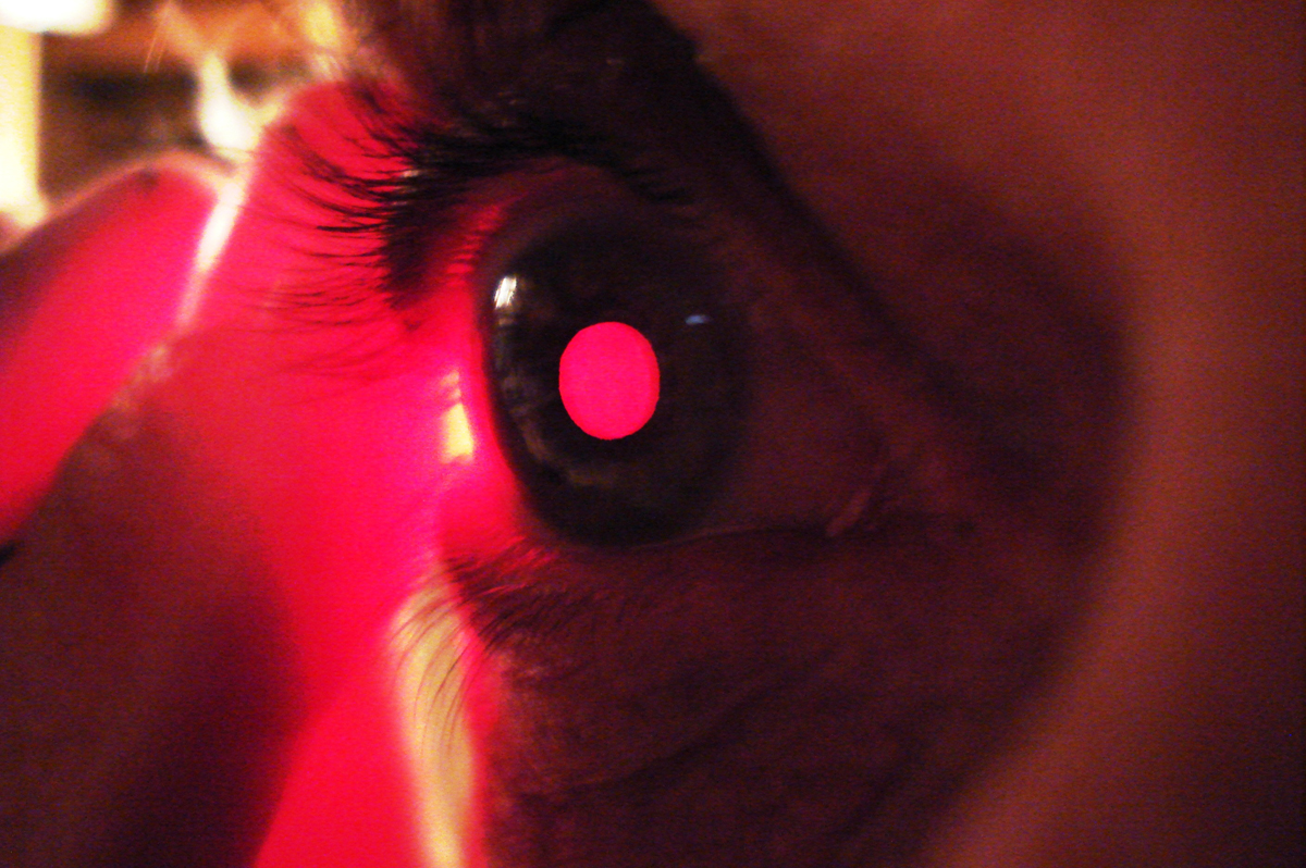
3. Stand in front of a mirror in a dimly lit room and hold the LED against the skin on the outside corner of your right eye while turning your gaze toward your nose. If you turn your head slightly to the right, you’ll be able to use your left eye to see the pupil of your right eye glowing red in the mirror. By changing the focus of your eyes, you may be able to see blood vessels magnified by the cornea and lens in the right eye. The largest of these vessels are about the size of a human hair.
The Light Swirler
The Light Swirler is a device for smoothly moving a bright light in your peripheral view to cast shadows of the blood vessels and tissue surfaces on top of your retina. Think of it as a kind of retinal scanner, revealing the unique pattern used in real retinal scanners for identification purposes. This technique was first described in 1819 using a handheld candle. Our device will be a scaled-down version of an instrument described in 1926, which used an incandescent bulb. Again, in our modern version, we’ll use an LED.
You can get a preview of what you’ll see with the Light Swirler by covering one eye and looking straight at a blank wall with the other eye while you rapidly flash a focused beam of light on the pupil from the side. Done correctly, you will see a branching pattern blinking on and off.
With the Light Swirler the view is considerably more dramatic and detailed — the shadows writhe around without the distracting blinking, and you can even see the moving shadow from the slight depression in the retina at the center of vision (the fovea) and the texture of the surface at the back of the eye.
To make it, you’ll mount a small lazy Susan between a putty knife and a ceiling fan pan box so you can rotate the box from the back. The pan box will contain the LED, switch, and battery box.
LIGHT SWIRLER MATERIAL & TOOLS
- PVC ceiling fan pan box and round cover
- Bracket used in electrical boxes for supporting light fixtures
- Lazy Susan, 3″, square Rockler #28951 rockler.com
- Putty knife, plastic, 4″
- Battery holder, 2×AAA RadioShack #270-398 radioshack.com
- Slide switch RadioShack #275-0406
- LED, white RadioShack #276-0017
- Nuts and screws, #6-32, 1″ (3), 1⁄4″ (9–11)
- Pipe strap (optional)
- Paper
- Tape, double-sided
- Soldering iron and solder
- Common hand tools
- Drill
- Hot glue gun and glue sticks
- Dremel rotary tool with sanding drum and cutoff wheel (optional)
1. Start by making the crank arm. I used an electrical box bracket and a #6 screw and nut to make a crank as shown above. This was handy because the bracket already had 2 of the holes and also had bends started in the right spots. Alternatively, you could use stiff pipe strap or another source of metal.
2. Mark holes in the pan box for mounting the crank and the lazy Susan, and mark trim areas and holes on the putty knife for the lazy Susan. Drill holes, cut trim areas, and assemble with #6 screws and nuts. I used the Dremel sanding drum for all trimming. You can cut off the ends of the screws with a Dremel abrasive cutoff wheel.
3. Now that we have the rotating mechanism, we can work on the electrical part. Cut and sand down the protrusions on the outside of the fan box — I used the Dremel sanding drum. Glue the battery box inside the fan box. Bend the slide switch tabs in slightly, and cut and drill the box to allow it to be mounted as shown. Drill a pair of holes in the side of the box for the LED and glue it in place with hot glue so it points up at about 45°.
4. Wire the switch, battery box wires, and LED together, taking care to get the polarity right (the longer lead on the LED is positive and goes to the red wire). For added protection of the LED, you can add a bit of pipe strap, either as a hood or as a holder. Additional hot melt glue can also be added around the LED.
5. Mount the fan box to the cover/lazy Susan assembly using two #6 screws and nuts. Use double-sided tape to stick a disk of paper to the box.
To operate the Light Swirler, turn on the LED, close one eye, and hold the assembly close to the other eye. Rotate or oscillate the light while looking straight ahead at the blank surface. You should try to hold the Light Swirler so that the light is visible at all times but never in your direct line of sight. The light is focused by your eye onto a part of the retina far from your center of vision. This results in what is essentially a bright light source within the eye itself, moving and shining on the blood vessels from a very oblique and unusual direction. The blood vessels lie a little above the light-sensing retina, and the moving light source never gives the retina time to compensate and cancel out the shadows.
The shadow image you see is unique to your eye. A real retinal scanner works by measuring the reflectance from a beam of infrared light as it sweeps a circular path on the retina. The changes in reflection due to intercepted blood vessels result in a unique digital signature.
The Brain Scanner
The Brain Scanner allows you to see the nerve activity on your retina, and because your retina is considered part of your brain, you will actually be seeing your own brain activity. This effect was first reported in 1825 and is called “the blue arc phenomenon.” It’s a subtle effect but I consider it the most beautiful thing to see in your eye.
BRAIN SCANNER MATERIALS & TOOLS
- Paper towel tube
- Aluminum can
- Cellophane, red
- Cellophane tape, matte
- Awl or knife
1. Cut a disk out of an aluminum beverage can to fit over the end of the paper towel tube, similar to how we made the Shadowscope.
2. Punch a small hole in the disk with an awl or the tip of a knife, and tape some red cellophane over the hole using matte-finish tape. Now tape the disk over the end of the cardboard tube as shown.
To use the Brain Scanner, hold it up to your right eye and line it up with a bright light source. Close both eyes for 30 seconds and then open the right eye and look to the right of the glowing red dot. You should see 2 blue arcs going from the red dot toward the right somewhat. They will fade after a few seconds, and if you look directly at the red dot they’ll disappear.
It may take several tries to see this. Between tries you need to open your eyes, then close them again for 30 seconds to partially accommodate them to the dark. You can use your left eye, but you must then look to the left of the red dot, and the arcs will be to the left.
The arcs are due to the activity of the nerves that are carrying the signal from the photoreceptors responding to the image of the red dot on your retina. Your retina is actually an outgrowth of your brain, so you’re seeing brain cell activity!
Conclusion
This article appeared in MAKE Volume 29, page 62.
There’s a lot of room for substitution and innovation on any of these projects — use your imagination and what you have available to save time and money.
For more information on floaters and the blue arcs, see the “Amateur Scientist” articles by Jearl Walker in the April 1982 and February 1984 issues of Scientific American. You can also view and download articles I wrote for The Physics Teacher and The American Biology Teacher, which describe these and other things you can see inside your eyes at makezine.com/go/mauser and makezine.com/go/mauser2.
This article appeared in MAKE Volume 29, page 70.


